TissueFAXS SPECTRA
Multispectral Tissue Cytometer
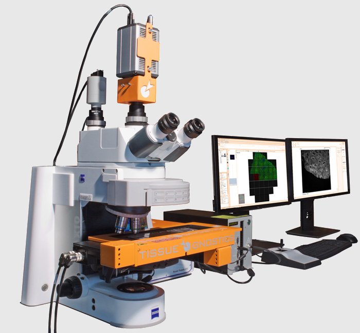
TissueFAXS SPECTRA systems provide Multispectral Imaging, Spectral Unmixing and Contextual Tissue Cytometry Image Analysis on the basis of the TissueFAXS platform.
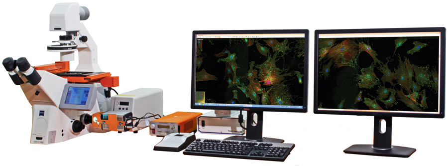
SPECTRA technology is available on any TissueFAXS system configuration, be it upright, inverted, TissueFAXS Histo, TissueFAXS Fluo, TissueFAXS PLUS or Slide Loader-based (SL).
TissueFAXS SPECTRA
UNIQUE FEATURES
- Multispectral fluorescence imaging
- Multispectral brightfield imaging
- 8 slides scanning
- Inverted or upright
- Slide ID scanner
- Quantitative image analysis option
- TMA & FISH/CISH scanning & analysis
- Automatic tissue detection
- Image and metadata storage, management and archiving
Technology
TissueFAXS SPECTRA is based on a liquid crystal tunable filter and the spectral unmixing engine of StrataQuest analysis software. Liquid crystal tunable filters are optical filters that use electronically controlled liquid crystal elements to transmit a selectable wavelength of light and exclude others. They can be tuned to specific wavelengths within the visible spectrum at high speeds. This makes them ideal for a quick building of the lambda stacks (a.k.a “Image Cubes” or “Spectral Cubes”), which then can be used for spectral unmixing.
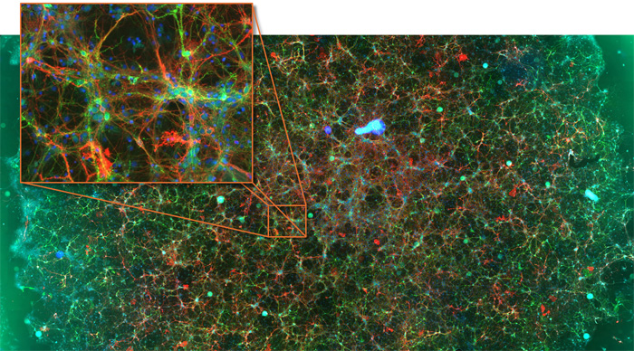
This permits the separation of multiple colors in both, brightfield and fluorescence. The following graphic shows the process from building the lambda stack to color deconvolution via the StrataQuest spectral unmixing engine. The result is four greyscale channel images corresponding to the four unmixed colours and an overlay image analogous to a fluorescence overlay image.
Contextual Tissue Cytometry
The separated channel images can then easily be used for contextual tissue cytometry analysis as provided by StrataQuest. This technology perfectly matches a current trend to look at multiple markers in parallel in order to better understand interactions among and between different cell types and cellular subpopulations.
PROPERTIES
Design
Modular (Microscope, Cameras, Light-source, PC with 2 Monitors)
Microscopy mode
Multi Spectral Fluorescence, Brightfield
Compatible Slide Formats
All standard and over-sized slides
Slide Capacity
8
Objectives
Up to 7 Objectives (2.5-100x)
FL-Channels
8
Camera
CMOS Camera (8/10-bit, 4.2 Megapixel, Color)
sCMOS Camera (8/16-bit, 4 Megapixel, Monochrome)
Light Source
Solid-state light engine
Tissue Microarray (TMA)
YES
Image Analysis Software
QUEST line (Algorithms for tissue detection, area measurement, scattergrams for cells and many more)
Supported Image Formats
TissueFAXS, OME-TIFF, TIFF, JPEG, BMP, PNG
Viewer
Integrated, stand-alone freeware

