Promicra QuickPHOTO
Deep Focus Module
Extended Depth of Field (EDF) Software
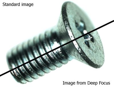
Deep Focus is a so called “focus stacking” or “z-stacking” or “EFI – Extended Focal Imaging” or “EDF – Extended Depth of Field” software module for QuickPHOTO programs. This module creates images, which are COMPLETELY IN-FOCUS, which cannot be achieved by a standard use of microscopes, because of their limited depth of field.
Deep Focus module can be used with stereomicroscopes as well as with other types of optical microscopes for observation in transmitted or reflected light. The module is also suitable for macro-imaging. The whole process can be automated using motorized focusing modules provided by PROMICRA and other suppliers.
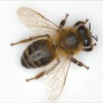
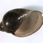
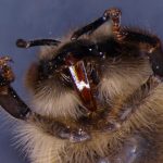
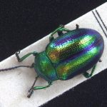
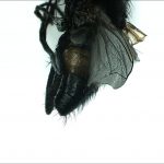
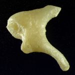
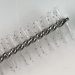
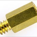
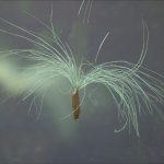
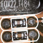
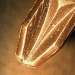
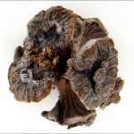
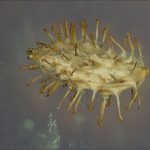
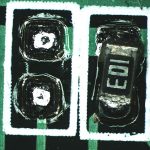
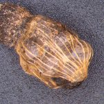
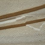
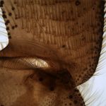
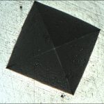
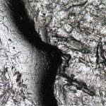
Gallery of EDF Images:
You can display the detail by clicking on the preview.
Images from Stereomicroscopes:
Overview:
Extended Depth of Field (EDF) Deep Focus, software module solves an age old imaging problem by finally being able to provide scientists and researchers the image they could only imagine, namely one that is COMPLETELY IN-FOCUS. The narrow depth of field associated with optical microscopy has always required microscopists to continually focus back and forth through the Z-axis of an object. If a picture was required, even selecting the “best picture” still left most of the image out of focus. Now, one can effortlessly acquire a series of partially focused images and quickly produce an Extended Depth of Field (EDF) picture. More than 100-times the original depth of field is possible!
Functions Overview:
- Universality – the module can be used with:
- Stereomicroscopes
- Optical microscopes for observation in transmitted light
- Optical microscopes for observation in reflected light
- Electron microscopes
- Macro images
- Excellent image quality suitable for publication – the module produces high quality images without artefacts
- Automation – the process of creating the images with extreme depth of field can be automated using optional module for z-axis motorization or a motorized stand or a microscope
- Can be used with HDR module to produce fully focused HDR images (EDF + HDR).
- Easy to use and user friendly graphical interface
- Options to compose images opened in the QuickPHOTO program as well as images stored on a hard drive
- Compatibility with QuickPHOTO CAMERA, QuickPHOTO MICRO and QuickPHOTO INDUSTRIAL programs in version 2.3 or higher
How It Works
Principle of Function:
1. Acquisition of a Stack of Slices with Different Planes of Focus
Because the depth of field of microscopes is limited due to optical principles (the higher the magnification, the shallower the depth of field is), a completely focused image needs to be composed from a series (stack) of standard images with different planes of focus, by software. A stack of several so called “slices” (images with different planes of focus) needs to be acquired first. Each of the slices contains different areas of a specimen in focus. The slices might be acquired manually by successive manual refocusing of a microscope or automatically using a PC controlled microscope z-axis motorization (see the Automation tab).
 The animation above displays sequentially the slices intended for composition acquired from a stereomicroscope and the image composed by Deep Focus module. Movement of the specimen in the field of view occurs as well as minor scale changes.
2. Composition of a Fully Focussed Image
The next step is a composition of a fully focussed image, from the stack of acquired slices, using the Deep Focus module. Only the areas, that are in focus, are used by the Deep Focus module from each of the slices for composition of a fully focused image. Possible specimen movements in the field of view and scale changes are compensated automatically during the composition process. The result is a high quality fully focussed image (see example bellow or visit the gallery of composed images).
Automation of the Composition Process
The whole process of acquisition of a stack of slices and composition of a fully focused image can be automated using PC controlled z-axis motorization of a microscope (using additional focus drive or motorized stand). The automation makes routine work with Deep Focus module much more comfortable.
The automated procedure is very straightforward. It consists of following steps:
- Focusing the microscope to the bottom (lowest) part of the specimen using the camera’s live view and setting the lower limit of the focus range.
- Focusing the microscope to the top (highest) part of the specimen using the camera’s live view and setting the upper limit of the focus range.
- Selecting the number of slices to be acquired or a step between them.
- Clicking the Start button to begin the whole process.
- Waiting a while until the fully focused image appears.
Gallery of Images from Upright Microscopes:
Transmitted Light:
Gallery of Images from Upright Microscopes:
Reflected Light:
System Requirements:
| Minimum Requirements | Recommended Specifications | |
| Processor | Single-core 2.4 GHz | Multi-core processor |
| Operating Memory | 1 GB | 2-4 GB |
| USB Ports | 2x USB 2.0/3.0 | 2x USB 2.0/3.0 |
| Operating System | Microsoft® Windows® XP(SP3)/Vista/7/8/8.1/10 |
Microsoft® Windows® 7/8/8.1/10 |
| Monitor Resolution | 1024 x 768 pixels | 1920 x 1080 pixels |

